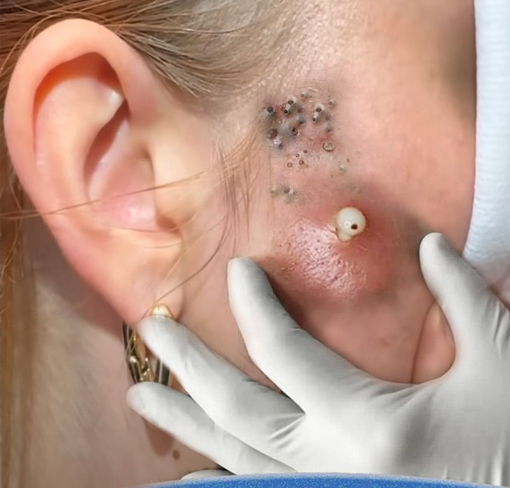
The image provided presents a compelling, complex dermatological case, highlighting two distinct and co-existing types of lesions in the periauricular (near the ear) and temporal region of the face. This scenario, featuring both numerous clustered blackheads and a large, inflamed pustule, offers a window into the multifaceted nature of acne and follicular disorders.
This article will explore the specific characteristics of these lesions, the potential underlying causes of their clustering and severity, and the specialized clinical procedures required for effective treatment.
Understanding the Dual Nature of the Lesions
The photograph clearly delineates two different acne forms developing simultaneously, indicating a varying response within the skin’s follicular structure.
1. The Cluster of Blackheads (Comedones)
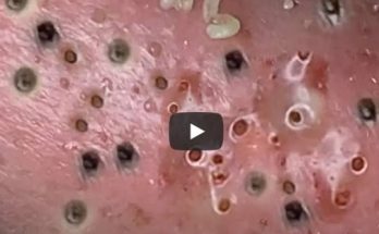
In the upper section of the image, there is a dense cluster of small, dark spots. These are open comedones, commonly known as blackheads.
-
Formation and Appearance: Blackheads occur when a hair follicle is clogged with a mixture of sebum (oil) and dead skin cells. Because the pore opening remains wide, the trapped material is exposed to oxygen, leading to the process of oxidation. This chemical reaction is what causes the material to turn dark brown or black, not dirt.
-
A Unique Pattern: Clustered Comedones: The high concentration and close proximity of these lesions suggest a unique and often challenging condition. This pattern can sometimes be associated with:
-
Acne Aestivalis (Mallorca Acne): Often linked to sun exposure combined with certain topical products (like heavy sunscreens or oils).
-
Acne Excoriée: While typically self-inflicted, the clustering might be an area of habitual picking.
-
Drug-Induced Acne: Certain medications can cause highly inflammatory and clustered lesions.
-
Follicular Hyperkeratinization: A localized area of the skin where the dead skin cells (keratinocytes) are produced and shed excessively, leading to rapid and severe clogging of multiple adjacent pores.
-
2. The Large, Inflamed Pustule/Nodule
Dominating the lower portion of the image is a large, raised, red, and swollen lesion with a distinct yellowish-white center. This is a severe form of inflammatory acne, likely a large pustule or a nodule that has come to a head.
-
Inflammatory Process: This type of lesion develops when the pressure from the trapped sebum, keratin, and bacteria (C. acnes) causes the follicle wall to rupture beneath the skin’s surface. The contents spill into the surrounding dermis, triggering a strong immune response.
-
Pus Formation: The body rushes white blood cells to the site to fight the bacteria, resulting in a localized accumulation of pus—the visible white/yellow center. The redness and swelling around the lesion indicate significant tissue inflammation.
-
The Risk: Due to its size and depth, this lesion carries a high risk of scarring (atrophic or hypertrophic) and post-inflammatory hyperpigmentation (PIH) if not treated carefully.
The Clinical Necessity: Professional Treatment and Extraction
Treating such complex and severe lesions requires a professional, multi-pronged approach, as attempting home extraction could lead to infection, deeper rupture, and permanent scarring. The gloved hand in the photo confirms that a clinical procedure is underway.
Treatment Strategy
-
For the Clustered Comedones (Blackheads):
-
Preparation: The skin is often pre-treated with topical retinoids (like tretinoin or adapalene) to normalize follicular keratinization, or prepared with steam and exfoliating solutions to loosen the plugs.
-
Extraction: A skilled clinician uses a sterile comedone extractor tool or a fine, sterile needle to gently press and remove the compacted material from each pore. The close clustering requires meticulous attention to avoid collateral damage to the surrounding skin.
-
-
For the Large Pustule/Nodule:
-
Incision and Drainage (I&D): For large lesions that have successfully come to a head (fluctuant), a small, sterile incision may be made to drain the pus and built-up pressure. This reduces inflammation and provides immediate relief.
-
Corticosteroid Injection: For deep, non-draining lesions (cysts/nodules), a dermatologist may inject a diluted corticosteroid solution directly into the lesion. This dramatically reduces inflammation and pain, often preventing the lesion from getting bigger and minimizing the risk of a severe scar.
-
Careful Extraction: If the lesion is deemed suitable for manual removal, the clinician uses slow, even pressure to ensure the complete evacuation of the follicular contents without pushing the material deeper into the skin.
-
Anatomical Consideration
The location of these lesions near the ear and temporal bone is also significant. The skin here can be relatively thin and less mobile compared to other facial areas, which can make extractions more sensitive and potentially more prone to visible bruising or trauma if not handled delicately. The proximity to the hairline also suggests a possible role of hair products or hair hygiene practices in contributing to the clogging.
Conclusion: A Case for Specialized Dermatological Care
The image serves as a powerful illustration of the complexity and variety of acne. It highlights a skin environment where severe inflammation and chronic clogging co-exist, demanding highly specialized care. While the visual spectacle of the extraction process is compelling, the underlying message is the need for appropriate diagnosis and professional, sterile procedures to safely manage and clear such intricate dermatological conditions, ultimately preserving skin health and minimizing long-term damage.
Would you be interested in learning about the different types of tools used by professionals to perform these extractions?