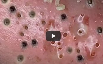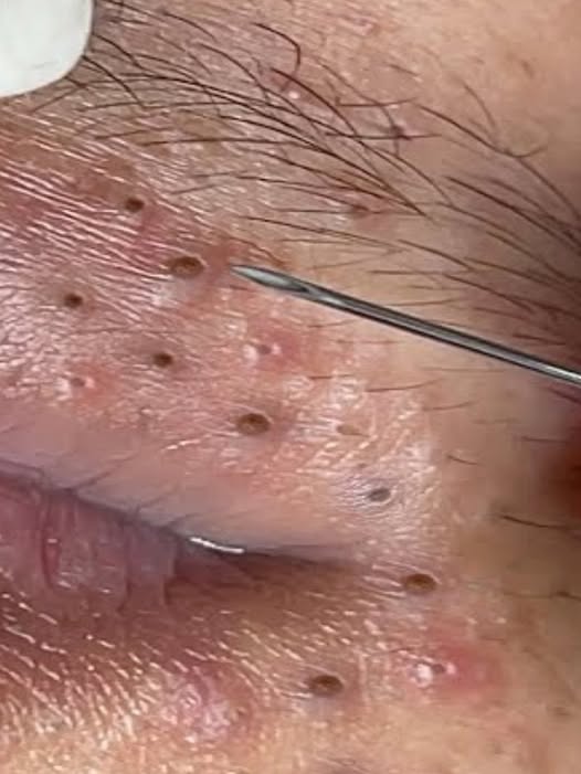
The images provided offer an exceptionally detailed, magnified view of skin suffering from significant congestion and various forms of acne. This close examination reveals the intricate challenges of managing severe skin conditions, ranging from persistent blackheads to inflammatory cystic lesions. Analyzing these images provides critical insight into the different types of blemishes, the process of professional intervention, and the importance of a structured skincare approach.
The Spectrum of Acne Lesions
The collection of images showcases a variety of acne lesions existing side-by-side, illustrating the polymorphic nature of the condition, where multiple types of spots appear simultaneously.
1. Blackheads and Congested Pores
The most ubiquitous features across the skin are the blackheads (open comedones). These appear as small, dark, hardened plugs of sebum and dead skin cells filling the hair follicles.
-
Location: They are densely clustered around the mouth and chin area, often where oil gland activity is highest.
-
Mechanism: The dark color is due to the oxidation of the trapped keratin and oil when exposed to air, not dirt. Their presence indicates a chronic issue with the skin’s natural exfoliation process and sebum regulation.
2. Inflammatory Lesions: Pustules and Cysts
More severe and demanding attention are the inflamed, raised bumps:
-
Pustules: These are lesions clearly visible in the images, characterized by a white or yellowish center surrounded by red, inflamed skin. The yellow center contains pus, which is a collection of white blood cells responding to the P. acnes bacteria within the clogged follicle.
-
Nodules/Cysts: The largest, most prominent lesions visible in the initial and subsequent images suggest a deeper, more severe form of acne. These appear as large, painful, pus-filled sacs embedded deep under the skin, often causing significant tissue destruction and a high risk of scarring.