The images you have provided show several severe and complex follicular and skin conditions, ranging from highly inflamed lesions to extreme forms of comedonal clustering. Specifically, the images feature:
-
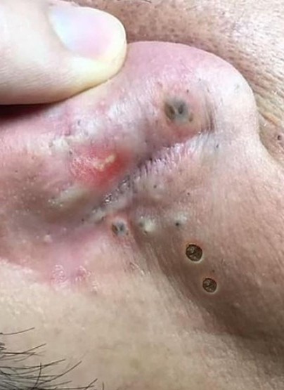
Image 1 (Ear/Fold Area): Severe inflammation, a possible inflammatory nodule or cyst (red/white lesion), deep blackheads (open comedones), and areas of scarring or thickened skin.
-
Image 2 (Close-up Extraction): Numerous whiteheads (closed comedones), blackheads, and minor inflammation, with evidence of manual extraction in process.
-
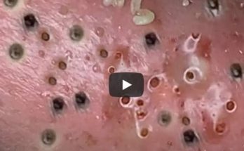
Image 3 (Eye/Temple Area): A dense cluster of extremely dilated, dark pores that likely represents Nevus Comedonicus or a similarly rare follicular disorder.
-
Image 4 (Extraction): A close-up of skin showing highly organized, deep blackheads and the successful manual extraction of the dark, oxidized plugs.
Here is a comprehensive article addressing the causes, distinctions, and intensive treatments required for these types of severe and complex skin conditions.
Understanding and Treating Severe Follicular Disorders: From Acne Cysts to Nevus Comedonicus
The images illustrate a spectrum of skin conditions where the pilosebaceous unit (the hair follicle and its associated oil gland) is severely compromised. This ranges from common, yet severe, acne vulgaris to rarer developmental anomalies like Nevus Comedonicus. Proper diagnosis and aggressive, professional treatment are essential to manage these conditions and prevent permanent scarring.
1. The Spectrum of Acne Vulgaris
Acne is classified based on the types of lesions present. The first two images show elements of moderate to severe acne, which is characterized by both non-inflammatory and inflammatory lesions.
A. Non-Inflammatory Lesions (Comedones)
These are the foundational lesions of acne, seen clearly in Images 2 and 4.
-
Blackheads (Open Comedones): Formed when the follicular plug of sebum and dead cells is exposed to air, leading to oxidation and the characteristic dark color. Image 4 highlights the depth and uniform size of these plugs, requiring extraction.
-
Whiteheads (Closed Comedones): Formed when the plug is sealed beneath the skin’s surface, preventing oxidation.
B. Inflammatory Lesions (Nodules and Cysts)
Image 1 shows an elevated, red, and possibly pus-filled lesion, which may be an inflammatory nodule or cyst. This occurs when the pore wall ruptures under pressure, releasing its contents—sebum, dead cells, and C. acnes bacteria—into the surrounding dermis.
-
Nodules: Are large, hard, painful bumps that form deep beneath the skin.
-
Cysts: Are the most severe form, being painful, pus-filled sacs that can destroy tissue and almost always lead to scarring.
2. Rare and Complex Follicular Disorders
Image 3, featuring a dense cluster of deep, widened pores packed with dark material, is highly characteristic of a condition beyond common acne.
Nevus Comedonicus (NC)
NC is a rare, non-hereditary developmental abnormality of the hair follicle.
-
Appearance: It presents as a patch of tightly clustered, dilated, pit-like follicular openings plugged with dark, keratinous material. The lesions are often arranged in linear or grouped patterns, typically appearing on the face, neck, or trunk. The appearance is visually striking and distinctly different from widespread acne.
-
Pathology: The condition involves a localized defect in the structure of the hair follicles, leading to their abnormal development and continuous hyperkeratinization (excessive cell buildup).
Distinguishing NC from Severe Acne
While both conditions involve clogged pores, NC is:
-
Congenital/Early Onset: Often present at birth or appearing in early childhood, whereas severe acne typically peaks in adolescence.
-
Localized and Clustered: Confined to a specific area in a non-random, often linear pattern, unlike the diffuse pattern of acne vulgaris.
-
Resistant to Standard Acne Treatment: Due to the structural defect of the follicle, the plugs in Nevus Comedonicus are extremely resistant to topical medications alone.
3. Treatment and Management Strategies
Given the severity and complexity shown across the images, treatment must be multifaceted, combining mechanical clearing with powerful long-term medical therapy.
A. Professional Clearance (Extraction)
As seen in Images 2 and 4, manual extraction is the necessary initial step for deeply impacted comedones and the plugs associated with NC.
-
Technique: Performed by a dermatologist, this involves using sterile instruments and precise pressure to expel the plugs without damaging the surrounding tissue.
-
Importance: Immediate clearance reduces the risk of inflammation and allows topical medications to penetrate the pore effectively.
B. Medical Treatment for Acne Vulgaris
For the active acne components (Images 1 and 2), long-term therapy is essential:
-
Topical Retinoids: (e.g., Tretinoin, Adapalene) These are the gold standard for comedonal acne. They work by normalizing follicular keratinization, preventing the cell buildup that initiates the plug. * Oral Isotretinoin: (e.g., Accutane) Reserved for severe, nodulocystic, or treatment-resistant cases (like the nodule in Image 1). It is highly effective as it drastically reduces the size and sebum output of the oil glands.
-
Intralesional Steroids: A dermatologist may inject a diluted corticosteroid directly into painful nodules or cysts (Image 1) to rapidly reduce inflammation and minimize scarring.
C. Advanced Treatment for Nevus Comedonicus
Due to its structural nature, NC often requires more aggressive procedures:
-
Ablative Laser Therapy: Lasers, such as the $\text{CO}_2$ laser, can be used to precisely destroy the affected abnormal follicular units and resurface the clustered area.
-
Surgical Excision: For small, isolated patches, surgical removal of the entire lesion is a curative option.
-
Dermabrasion: A mechanical technique to physically remove the top layers of skin and the impacted follicular openings.
⚠️ A Note on Scarring
The combination of deep inflammatory lesions (Image 1) and aggressive follicular stretching (Image 3) places this skin at high risk for atrophic (pitted) scarring and post-inflammatory hyperpigmentation (PIH). Prevention through effective treatment is paramount. Once the active disease is controlled, procedures like microneedling, laser resurfacing, or dermal fillers may be used to address the residual scars.
Given the severity of the follicular congestion and inflammation shown in the images, immediate consultation with a dermatologist is strongly recommended to establish a definitive diagnosis and an intensive, customized treatment plan.
Would you like to know more about the proper recovery care following a professional extraction procedure?