The two images presented offer a vivid, clinical look into the most challenging manifestations of acne vulgaris, showcasing both dense clustering of non-inflammatory lesions and the intense inflammation of cystic acne. These visual examples—one near the temple/ear, the other on the cheek—highlight the complexity, severity, and diverse pathology that can occur within the same skin condition.
This article will break down the types of lesions observed, explore the underlying causes of this severity, and discuss the specialized dermatological interventions necessary for treatment.
🌑 Image 1: The Juxtaposition of Inflammatory and Comedonal Acne
The first image captures a section of skin near the temple and ear, demonstrating a striking contrast between a massive, acute inflammatory lesion and a field of chronic non-inflammatory ones.
1. The Solitary, Inflamed Pustule/Nodule
A large, raised, erythematous (red) lesion with a distinct, prominent yellowish-white center dominates the scene.
-
Pathology: This is a severe form of inflammatory acne, likely a large pustule or an active nodule. It forms when the wall of a hair follicle ruptures deep beneath the surface, spilling sebum, keratin, and C. acnes bacteria into the surrounding dermis.
-
Appearance: The resulting intense inflammation and immune response lead to the accumulation of pus, which is visibly pointing (or “coming to a head”) at the skin’s surface. Its size indicates significant tissue involvement and pain, carrying a very high risk of permanent scarring (atrophic or hypertrophic).
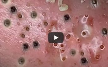
2. The Clustered Microcomedones and Blackheads
Adjacent to the large lesion is a dense patch of small, dark spots scattered across the skin.
-
Pathology: These are primarily open comedones (blackheads). The dark color is due to the oxidation of the trapped sebum and cellular debris exposed to the air. The small size suggests they may be new or chronic microcomedones that are close to the surface.
-
The Cluster: The tight grouping of these lesions is often an indicator of severe follicular hyperkeratinization, where the skin cells lining the follicle are shed and stick together too rapidly, leading to the simultaneous clogging of multiple adjacent pores.