⚠️ Complex Dermatological Cases: A Visual Analysis of Advanced Skin Lesions and Clinical Intervention
The three provided images depict a range of severe and complex dermatological conditions that are clearly under professional care. The photographs capture different stages of skin disease and active clinical procedures, underscoring the critical need for expert diagnosis and intervention beyond routine skincare.
🔬 Image Analysis: A Spectrum of Skin Pathology
Image 1: The Atypical Pigmented Lesion
This image presents a close-up of a patient’s temple/cheek area, highlighting a particularly worrisome spot:
-
Atypical Lesion: A distinct, dark, central lesion surrounded by a noticeable halo of blue-gray discoloration (perilesional pallor/haloing). The center appears crusted or ulcerated.
-
Differential Diagnosis: The irregular color, unique halo effect, and crusted texture necessitate a thorough evaluation. While it could represent an atypical form of a deep inflammatory lesion or a resolving viral wart, it raises concerns for conditions such as:
-
Deep Melanoma or Pigmented Basal Cell Carcinoma (BCC): The blue-gray hue can indicate pigment deep in the dermis.
-
Atypical Nevus (Mole): A mole undergoing change or inflammation.
-
-
Surrounding Comedones: Multiple smaller, dark spots (blackheads or keratosis pilaris-like lesions) are clustered around the brow and temple, indicating general follicular blockage or sun damage in the area.
-
Clinical Context: A gloved finger is seen pointing at the lesion, confirming professional examination.
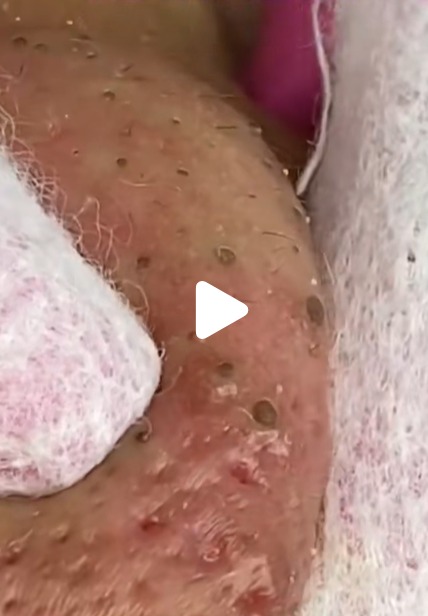
Image 2: Severe Inflammatory and Crusted Acne
This photograph shows a highly inflamed area, likely the forehead or cheek, characteristic of severe acne vulgaris:
-
Nodulocystic Acne: The general redness and swelling (erythema and edema) indicate deep, severe inflammation consistent with nodular or cystic acne.
-
Active Pustules: Several yellowish-white, raised spots contain pus, showing active infection and inflammation within the follicles.
-
Crusted/Oxidized Lesions: The most striking feature is the clusters of large, dark-brown to black, raised lesions. These are likely:
-
Heavily Oxidized Macro-comedones (Giant Blackheads): Extremely large, long-standing plugs.
-
Post-Rupture/Post-Procedure Crusting: Scabs forming over sites where deep cysts or pustules have either ruptured or been professionally drained/extracted.
-
-
Intervention: The presence of protective goggles and a probe or optical fiber connected to a medical device suggests the patient is undergoing a light-based therapy (e.g., Photodynamic Therapy, IPL, or laser treatment) aimed at reducing inflammation and targeting the P. acnes bacteria.
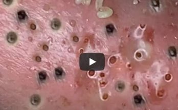
Image 3: Congested Skin and Aggressive Extraction
This image is a close-up of a technique being performed on a highly congested area:
-
Pore Congestion: The skin is visibly red and swollen with numerous small, dark spots, indicating widespread comedones and sebaceous filaments.
-
Extraction in Progress: A gloved hand (partially visible) and a white cloth/gauze are being used to manipulate the skin. The presence of large, empty-looking, distinct circular openings (similar to the first prompt’s image) suggests the procedure is a manual extraction aimed at clearing the follicular plugs.
-
Risk: The degree of inflammation and the visible manipulation suggest a potentially aggressive extraction technique, which, while effective for clearing plugs, carries a significant risk of post-inflammatory hyperpigmentation (PIH) and scarring if not performed carefully in a sterile setting.
⚕️ The Necessity of Dermatological Care
These combined images illustrate that these skin conditions are beyond the scope of routine cosmetic care and require medical-grade intervention.
1. Accurate Diagnosis and Safety
The atypical lesion in Image 1 is a prime example of why self-diagnosis is dangerous. A lesion with such concerning features requires immediate evaluation, possibly including dermoscopy and a biopsy, to rule out skin cancers.
2. Systemic Disease Management
Severe cystic acne (Image 2) is a chronic inflammatory disease that can lead to permanent scarring. Effective management often requires oral medications (such as isotretinoin or oral antibiotics), which must be prescribed and monitored by a dermatologist. Topical treatments alone are insufficient for such deep, widespread inflammation.
3. Minimizing Scarring and Complications
The aggressive nature of the required treatments (extractions, lasers, deep drainage) must be balanced with protocols to prevent long-term damage. Dermatologists use sterile tools, perform procedures like intralesional steroid injections to rapidly reduce deep cyst inflammation, and guide patients through post-procedure healing to minimize the risk of permanent atrophic (pitted) scarring.
In summary, these photographs serve as a powerful reminder that severe acne and atypical pigmented lesions are serious medical concerns. They highlight the specialized nature of dermatological practice, where advanced diagnostics and procedural skills are crucial for preserving the patient’s health and skin integrity.
Would you like to learn more about the signs that indicate a skin lesion should be immediately checked by a doctor?