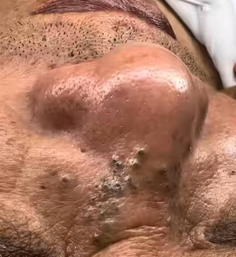
The uploaded image presents a close-up view of the skin, primarily focusing on the area around the nose and adjacent facial regions. This visual evidence showcases a constellation of skin features, some of which appear significantly raised and inflamed, while others are characterized by a darker, potentially porous texture. A thorough examination of these features can provide insights into potential dermatological conditions and the importance of professional care.
The Prominent Nasal Feature
The most immediately noticeable element in the image is the significantly enlarged and bulbous appearance of the nose. The skin over this area appears taut, shiny, and erythematous (reddened), suggesting underlying inflammation, swelling, or structural changes.
-
Potential Condition: Rhinophyma. This is a severe form of rosacea, often classified as Phymatous Rosacea. Rhinophyma is characterized by a gradual enlargement of the nose due to the overgrowth of sebaceous (oil) glands and connective tissue. It gives the nose a swollen, red, and lobulated appearance. While the exact cause is unknown, it is associated with long-term, untreated rosacea. The visual presentation in the image—the enlarged, rounded shape—is highly suggestive of this condition.
-
Other Considerations: Less commonly, this kind of swelling could be related to severe, localized infections, deep cysts, or other rare skin tumors, though the overall texture and presentation lean strongly toward a chronic condition like rhinophyma.
Features Below and Adjacent to the Nose
Below the main structure of the nose, and possibly extending onto the cheek area, the skin exhibits several other distinct features that merit discussion:
1. Numerous Small, Dark Lesions
A cluster of small, dark, raised lesions is visible in the lower part of the nasal/perinasal area. These features often have a specific appearance:
-
Blackheads (Open Comedones): Many of the small, dark spots could be open comedones. These form when a hair follicle is clogged with sebum (oil) and dead skin cells. The ‘black’ color is not dirt, but rather the oxidation of the trapped material (melanin) when exposed to air. Given the close proximity to a potentially hyperactive sebaceous gland environment (as suggested by rhinophyma), the presence of numerous comedones is plausible.
-
Sebaceous Filaments: These are similar to blackheads but are a natural, often lighter-colored collection of sebum and dead cells that line the sebaceous gland duct. In skin with very active oil glands, they can appear enlarged and more noticeable. The dark color in the image, however, suggests they are likely true blackheads.
2. Scattered Papules and Pustules
Interspersed among the dark lesions are smaller, raised bumps, some of which appear to be skin-colored or slightly inflamed.
-
Acne Vulgaris: The presence of both comedones and inflamed bumps (which could be papules or small pustules) is a hallmark of acne. This suggests a concurrent inflammatory process in the skin’s oil glands.
-
Inflammatory Rosacea: Given the suspicion of rhinophyma, the smaller, inflamed bumps could also represent the papulopustular form of rosacea, which is often mistaken for acne. These lesions typically lack the true comedones (blackheads/whiteheads) characteristic of standard acne, although both can certainly coexist.
The Importance of Professional Dermatological Evaluation
It is crucial to emphasize that this analysis is based solely on the visual evidence provided in a single photograph and does not constitute a medical diagnosis. The complex nature of the features presented underscores the necessity of professional medical intervention.
-
Accurate Diagnosis: A dermatologist can distinguish between rosacea (including rhinophyma), severe acne, cysts, and other less common dermatoses through a clinical examination, and potentially with procedures like a skin biopsy.
-
Treatment Pathways:
-
Rhinophyma: Treatment for significant rhinophyma typically involves procedures to reshape the nose, such as dermabrasion, laser resurfacing, or scalpel excision to remove the excess tissue. Medications like oral isotretinoin may be used to slow the progression or reduce the size of the sebaceous glands.
-
Acne/Comedones: These are often managed with topical retinoids, benzoyl peroxide, and/or chemical peels. For severe cases, oral antibiotics or isotretinoin may be necessary.
-
Inflammatory Lesions: If part of rosacea, treatment would focus on reducing inflammation with specific topical or oral medications (e.g., metronidazole, azelaic acid, or doxycycline).
-
Conclusion: A Case for Comprehensive Skin Health
The photograph serves as a powerful visual example of chronic, severe, and potentially long-standing skin issues. The combination of significant dermal overgrowth on the nose and numerous follicular plugs and inflammatory lesions nearby suggests a complex dermatological picture, likely involving a severe form of rosacea with associated acne or follicular prominence. Seeking the care of a board-certified dermatologist is the essential next step to receive an accurate diagnosis, formulate an effective, personalized treatment plan, and manage the long-term health and appearance of the skin. Early intervention, especially in conditions like rhinophyma, can significantly impact the final cosmetic and functional outcome.
Would you like to know more about the general treatment options available for rhinophyma or severe acne?